Neurodegenerative diseases often involve the accumulation of misfolded proteins, that cells fail to refold or degrade and that eventually form aggregates. Among these, oligomers, small still-soluble aggregates, are the most toxic and can lead to neuronal death. Identifying the aggregation-prone regions of these proteins is key for developing new therapeutic approaches.
In over 97% of amyotrophic lateral sclerosis cases, TDP-43 (TAR DNA/RNA-binding protein 43) condensates accumulate in the cytoplasm. The carboxy-terminal region of TDP-43 is processed into smaller aggregation-prone fragments that are included into these condensates.
Akira Kitamura and their colleagues at Hokkaido University explored these fragments by expressing TDP-43 deletion constructs in murine neuroblastoma cells (N2A) and C. elegans. They found that TDP25, a fragment containing a glycine-rich domain and part of the RRM2 RNA-binding domain, is necessary and sufficient for condensate formation. The glycine-rich region drives this process, while the remaining N-terminal fragment is highly aggregation-prone. Only some of these condensates were stained by Amytracker 680, and therefore had an amyloid-like structure. This suggests that multivalent interactions and conformational changes in biomolecular condensates might facilitate the formation of amyloid aggregates of TDP-43.
Expressing TDP25 caused cell death in N2A cells and reduced lifespan in C. elegans, suggesting this fragment contributes to the neurotoxicity observed in amyotrophic lateral sclerosis. Therefore, stabilising TDP25 could be a potential therapeutic target to prevent toxic TDP-43 oligomers.
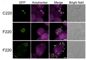
Image: Confocal images of N2A cells expressing different GFP-tagged TDP-43 carboxy-terminal fragments (green) stained with Amytracker 680 (magenta). In cells expressing the F220 fragment, not all the condensates formed are positive for Amytracker staining, suggesting that only a fraction of them contains amyloid fibrils. White arrows and yellow arrowheads represent the positions of Amytracker-positive and negative condensates in the cytoplasm. The asterisk in the images represents the nucleolus. Scale bar = 5 μm. Image from Figure 3E by Kitamura, A. et al. (2024) Communications Biology Chemistry, 7(1):743(CC BY 4.0).
Read More:
- Kitamura, A. et al. (2024) Hetero-oligomerization of TDP-43 carboxy-terminal fragments with cellular proteins contributes to proteotoxicity. Communications Biology, 7(1), 743
- Cascella, R. et al. (2023) An in situ and in vitro investigation of cytoplasmic TDP-43 inclusions reveals the absence of a clear amyloid signature. Annals of Medicine, 55(1), 72–88