Utricularia species, also known as bladderworts, are aquatic carnivorous plants. They have evolved one of the fastest movement mechanisms in the plant kingdom that allows them to trap and consume zooplankton, tiny insects, or algae. When an unsuspecting prey touches the sensory hairs on the trap door the bladder instantly generates negative pressure, sucking in water along with the prey. Scientists are still uncertain about the mechanism of resetting its trap. Some suggest that internal glands actively absorb and expel water, while others propose that specialized external glands play a crucial role in water expulsion.
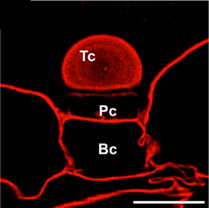
A study published in the International Journal of Molecular Sciences takes a deeper look into the external glands. By analyzing - using Carbotrace 680 - the structural layers of their cell walls, researchers have uncovered variations in homogalacturonans and hemicelluloses. These differences could be the key to understanding how bladderworts efficiently reset their traps and prepare for their next meal. The team found that the outermost layer of the gland cells of Utricularia dichotoma lacked arabinogalactan proteins, while the deeper layers were rich in them. The presence of these proteins correlates with the presence of hemicelluloses, which regulate cell wall expansibility and cell-to-cell adhesion, and suggests a functional specialization in water transport and secretion. This innovative use of Carbotrace highlights its potential for advanced plant biology research, helping scientists better understand how plants regulate fluid movements through cell wall composition.
This research not only sheds light on the biomechanics of one of nature’s fastest predators but also offers valuable insights into plant cell wall specialization and fluid transport.
Image: The terminal cell (Tc), pedestal cell (Pc), and basal cell (Bc) of the external gland in Utricularia dichotoma were stained with Carbotrace 680 (in red). The lack of staining in the latera walls of the pedestal cell (Pc) and the cell wall of the terminal cell (Tc) is probably related to the presence of cutin in these regions. Scale bar: 10 µm. Image adapted from Figure 6A by Płachno, B. et al. (2024) nternational Journal of Molecular Sciences, 25(23), 13124. (CC BY 4.0).